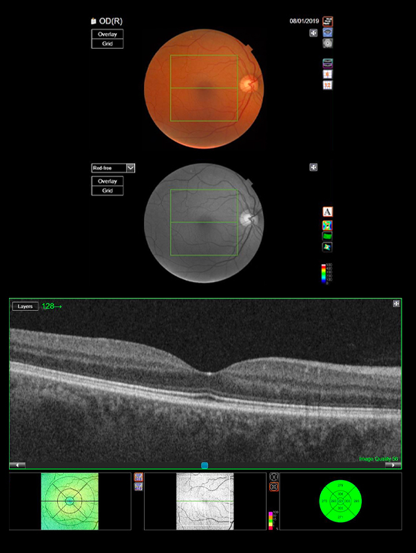
What is an OCT Scan?
OCT stands for Optical Coherence Tomography scan. It is a detailed scan using state of the art technology to help build up a 3D image of the eye.
By using such technology the team at Dipple and Conway are able to fully assess the health of your eyes. The scan allows us to really get a clear indication of what is happening below the surface in great detail by evaluating the different layers of the eye. The 3D imaging taken during the scan is actually made up of several individual scans, allowing us to evaluate each layer of the eye’s tissue with precision.
These scans can help to detect sight-threatening eye conditions. They are particularly useful for detecting conditions that would have otherwise gone unnoticed, until they started to affect your vision. Generally the earlier eye conditions are noticed, the easier they are to treat.
Did you know? Glaucoma can actually be detected up to four years earlier using an OCT Scan compared to an ordinary sight test.
Estimated Read Time: 11 minutes

Matthew Conway
CEO/Director
Published:
Updated:
In this article...
What areas of the eye does an OCT Scan focus on?
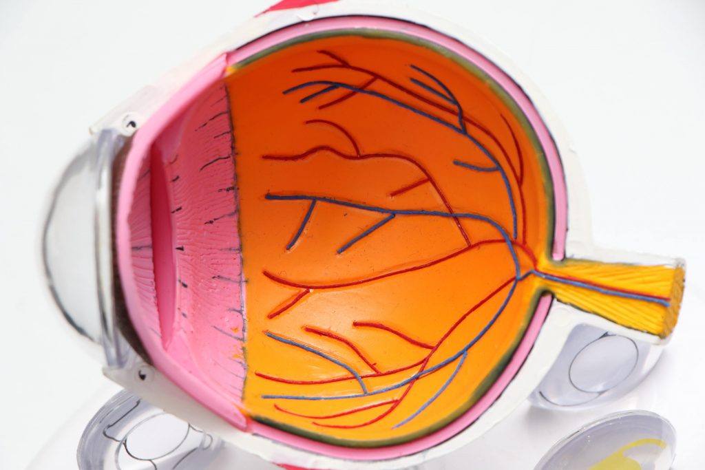
During your OCT Scan, our clinical assistants will take imaging of the retinal layers of your eye. This eye examination will guide our optometrists who will then study the scans and assess several parts of your eye, primarily your macula, your optic nerve and your cornea.
The Macula
Your macula is the part of your eye you need for clear central vision. It allows you to see fine detail. Without your macula functioning fully you would struggle to see well enough to read and would find it difficult to recognise faces.
The Optic Nerve
Your optic nerve is made up of over a million nerve fibres and forms a pathway which transmits information between your eye and your brain. Sensory information received by the eye is passed to your brain by way of electrical and chemical impulses. Should you experience optic nerve degeneration and/or damage, this could potentially cause distortion or loss of vision and, in a worse case scenario, blindness. Early detection is key in preventing this.
The Retina
The retina contains essential photoreceptor cells that capture the images/light that you see. The information collected is then transmitted via the optic nerve to the brain, where it is processed. There are many layers to the retina, with each layer having an essential function. OCT imaging allows us to detect signs of degeneration by viewing each layer individually and in detail.
At the very front of the eye sits the Iris and the cornea, which allow controlled and safe passage of this light to your retina.
What conditions do we look for in different parts of the eye?
As previously discussed, an OCT Scan provides us with a picture of three main areas within the eye, the macula, the optic nerve and the retina. The OCT examination allows us to see changes happening within these areas early on. Each section of the eye can present its own unique challenges and can be affected by different types of damage or disease, which is why it is so important to have a trained professional to take the time to assess your results.
At Dipple and Conway we never rush this process and the interpretation of this information is always performed by our highly trained optometrists who will evaluate the scans taken, and check each of the different layers of the eye to check for signs of degeneration or change.
Conditions that can be detected on an OCT Scan include;
Glaucoma
In short, Glaucoma is a degeneration of the optic nerve and is one of the main conditions that we look for in an OCT Scan. As this is one of the more serious issues we have gone into more detail on this condition further on in this article.
Swelling and bleeding from leaking vessels for example, in Diabetic Retinopathy
Diabetic Retinopathy can lead to vision loss and can develop in those who have type 1 or 2 diabetes. This is caused by damage to the blood vessels at the back of the eye, which is more likely to happen the longer someone has diabetes and if blood sugar levels aren’t controlled. This is because having too much sugar in your blood can cause blockages to the blood vessels that go to the retina, these blockages prompt new blood vessels to grow but these usually don't grow properly and often leak, causing the damage.
Not everyone will develop symptoms straight away, but once the condition develops symptoms may include blurred vision, dark areas in vision, floaters or even vision loss. It is important that if any of these symptoms are developed an optician should be seen as soon as possible.
Vitreous degeneration/Traction
This is an age related condition which tends to occur after the age of around 50. At this time of ageing the fibres that attach the vitreous to the retina shrink and pull away. During this process the vitreous sometimes doesn’t detach itself properly and remains stuck, the vitreous then pulls to try and detach itself from the macula which can lead to damage occurring.
Detached Retina
The retina can detach due to the jelly in your eye changing and if your retina becomes detached it is vital that this is treated quickly to prevent sight loss.
Symptoms to look out for include blurry vision, floaters suddenly appearing or increasing in numbers, dark shadows moving across your vision or flashes of bright light.
Macular hole
A macular hole is a small gap that opens in your macular (a small part of the retina). This part of the eye is used for more focused tasks such as reading and because symptoms include blurred or distorted vision reading can become difficult.
After some time this distorted vision may evolve into a black patch in your vision. This condition doesn’t lead to a full loss of sight, but it does require surgery to repair the hole. This condition most commonly affects those aged 60-80 and usually affects women more than men. Although the surgery will recover some of their vision, after surgery it will never return completely to normal.
Age related Macular Degeneration
Also known as AMD, this condition is usually found in those aged 50-60 and is a condition that affects the middle part of your vision. If left untreated vision can get worse but it does not result in full vision loss.
The first symptom found is most commonly blurred or distorted vision and as the condition gets worse there may develop a black spot in the centre of vision. Alongside these there are less common symptoms to look out for that include hallucinations or colours seeming dimmer than usual.
Central Serous Retinopathy
Fluid building up in the retina is the cause of this condition, this fluid comes from the choroid which is a layer of tissue.
This is more likely to develop in men in their 30s-50s. Symptoms may include distorted central vision, a dark spot in the centre of the eye, brown tinges on white objects and straight lines appearing bent or wavy.
What are the main conditions that can affect the macula?
A macular hole is, as it sounds, a small hole that appears in the macular. As the macular is responsible for your central vision, having a macular hole can affect your vision by causing blurriness, patchy vision and/or distortion. This is usually noticed when performing fine detail tasks such as reading or sewing.
Whilst a macular hole can be caused by injury, the most common cause of a macular hole is aging. Inside our eyes we have a jelly known as Vitreous Humour, which functions to support the shape of our eyes. As we age this jelly can break down and separate from the retina itself. This is common and can often happen without too much impact on your vision. Sometimes, however, when the jelly breaks down it does not happen smoothly. In this case, strong attachments between the jelly and the retina are broken with force. This in turn causes damage to the macula and a hole can form.
Another condition detectable with OCT scanning is the formation of an epiretinal membrane, sometimes referred to as Macular Puckering. This condition is caused when a thin layer of tissue on the surface of the retina (often the macula area) develops a wrinkled appearance. This condition can affect our vision and sometimes requires treatment.
What are the main conditions that can affect the optic nerve?
The optic nerve being in top condition is an essential factor in maintaining good eye health. One of the main conditions we look for during OCT imaging of the optic nerve, is Glaucoma.
Glaucoma is a degeneration of the optic nerve and is often associated with an increase in internal pressure within the eye, but not always. Although it is rare, Glaucoma can appear rapidly, ordinarily however the condition develops gradually.
If not diagnosed early the condition will often, eventually, lead to progressive loss of peripheral vision. In the early stages of Glaucoma, many cases actually carry no symptoms at all. As it progresses you may begin to notice your vision is slightly blurred, or you may see “halos” or “rainbows” around bright lights. If left untreated, Glaucoma can lead to complete loss of vision.
As aforementioned however, a properly assessed OCT Scan can help to diagnose Glaucoma up to 4 years earlier, and that is just one of the reasons that we believe they are a crucial factor in detecting anatomical anomalies and early warning signs.
Aside from checking for signs of Glaucoma, during an OCT Scan our optometrists will also check for signs of damage or other optic nerve problems, as it can also be affected by trauma, cardiovascular or demyelinating conditions and other external factors such as toxins.
How long does an OCT Scan take?
An OCT Scan will be carried out at the same time as your routine eye test. It will only take a few seconds to perform the procedure, thanks to our specialist equipment. The optometrist will then take some time to fully study and interpret the results. The findings will then be explained to you during your time spent with us.
Does it hurt?
No, the OCT Scan does not hurt as it is completely non-invasive. The technology we use doesn't even touch your eyes.
To get the best results, your optometrist may add some drops to your eyes to allow your pupils to dilate. This widening allows us to capture a clearer OCT image. This process can temporarily make your eyes more sensitive to light but it won’t be painful. In most cases, this part of the procedure is not necessary.
Is it safe to have an OCT Scan?
Yes, it is a non-invasive, safe procedure. In simple terms we are just taking a picture of the back of your eye. Just like how an ultrasound captures images, an OCT functions in a similar way, but rather than using sound waves as an ultrasound does, it uses a low powered laser and gathers the information using light.
How often do I need an OCT Scan?
We recommend that you have the scan alongside your routine eye test, which is preferably annually but at least every two years providing your eyes are healthy. By having an OCT Scan, even when your eyes seem healthy, we are able to keep an image of your eyes on file. By comparing your base scan with those taken in the future, it becomes easier to spot any changes within the eyes over time, and helps us to swiftly treat or monitor any issues that could lead to further eye damage.
Does everyone need an OCT Scan?
We recommend OCT Scans to all adults. They are particularly important for those who have a family history of eye disease such as Glaucoma as it allows us to pick up on signs of this nice and early.
The team here at Dipple and Conway are always on hand should you have any further questions, so feel free to give us a call or visit your local branch. As experts in eyecare for over 100 years, you can rest assured that your eyes will be looked after by the team here at Dipple and Conway, book your eye test and OCT Scan today.
Posted By
Matthew Conway
CEO/Director